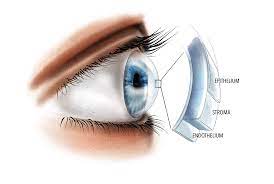
Corneal dystrophies are a rare group of genetic eye diseases. In corneal dystrophies, abnormal material accumulates in the cornea (the clear front window of the eye). The majority of corneal dystrophies affect both eyes. They spread slowly and in families.
The cornea is made up of five layers:
- The epithelium is the cornea’s outermost protective layer.
- Bowman’s membrane is a strong second protective layer.
- The stroma is the cornea’s thickest layer. It is composed of water, collagen fibres, and connective tissue. This strengthens the cornea while also making it more flexible and transparent.
- Descemet’s membrane: a protective inner layer that is thin and strong.
- Endothelium: the innermost layer of cells that drains excess water from the cornea.
Corneal dystrophies are caused by the accumulation of foreign material in one or more of the cornea’s five layers. The material may cause the cornea to become opaque. This can result in vision loss or blurred vision.
There are over 20 distinct types of corneal dystrophies. They are generally classified into three types:
- Dystrophies of the anterior or superficial cornea. These affect the cornea’s outermost layers, the epithelium and Bowman’s membrane.
- Stromal corneal dystrophies affect the stroma, the cornea’s middle and thickest layer.
- Posterior corneal dystrophies affect the cornea’s innermost layers, the endothelium and the Descemet membrane. Fuchs’ dystrophy is the most common type of posterior corneal dystrophy.
What are the signs and symptoms of corneal dystrophy?
The symptoms of corneal dystrophy vary depending on the type. Some people have no symptoms. In others, material buildup in the cornea causes it to become opaque (not clear). This results in blurred or lost vision.
Many people suffer from corneal erosion. This occurs when the layer of cells on the cornea’s surface (the epithelium) separates from the layer beneath (Bowman’s membrane). Corneal erosion causes: mild to severe eye pain, light sensitivity, and the sensation that something is in the eye.
Who is at risk of developing corneal dystrophy?
Because most corneal dystrophies are inherited, having a family history of the disease raises your risk.
Corneal dystrophies can manifest themselves at any age. Except for Fuchs’ dystrophy, most corneal dystrophies affect men and women equally. Women are more likely than men to be affected by Fuchs’.
Diagnosis
Your ophthalmologist will examine your eye if they suspect you have corneal dystrophy. They will also inquire about any eye disease in your family history.
Your ophthalmologist will use a slit lamp microscope to shine a thin, bright sheet of light into your eye. This allows the doctor to examine the front part of your eye thoroughly.
Even if a person has no symptoms, a routine eye exam may reveal that they have corneal dystrophy. In addition, genetic testing can detect corneal dystrophy in some cases.
What is the treatment for corneal dystrophy?
Treatment for corneal dystrophy is determined by two factors: the type of dystrophy and the severity of symptoms.
If you have no symptoms, your ophthalmologist may closely monitor your eyes to see if the disorder is progressing. In other cases, eye drops, ointments, or laser therapy may be necessary.
Recurrent corneal erosion is joint in people with corneal dystrophy. Antibiotics, lubricating eye drops, ointments, or special soft contact lenses that protect the cornea may be used to treat this condition.
If the erosion persists, other treatment options may include laser therapy or a scraping technique for the cornea.
A corneal transplant (also known as keratoplasty) may be required in more severe cases. The damaged or unhealthy corneal tissue is removed and replaced with clear donor corneal tissue. A partial cornea transplant (or endothelial keratoplasty) is used to treat endothelial dystrophies such as Fuchs’ dystrophy.

