The study of the cells that are extracted from fluids or bodily tissues is called cytopathology or cytology. It is carried out by healthcare providers in various settings. However, one of its most common uses is cancer screening and diagnosis.
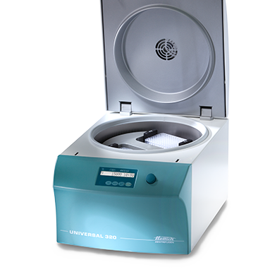
Using a cytology centrifuge is common in research and immunohematology laboratories. A cytology centrifuge is also a staple in blood stations, hospitals, chemical laboratories, and plasma stations.
How Cytology Centrifuges are Used
To prepare a cytology centrifuge smear, a funnel assembly will be mounted on the front of the microscope slide. Filter paper will be used to wipe and absorb any excess fluid on the funnel’s surface. To ensure the preservation of the cellular structure, the assembly will be placed in a cytocentrifuge that operates at 600—800 x g.
The centrifugal force will push the fluid through the funnel, concentrating the cells in a small area of the slide. The centrifugation process will also concentrate cells around twenty-fold, creating a one-cell-thick monolayer. This allows the evaluation of cellular morphology. The slide can be stained and fixed after.
The Different Applications of a Cytology Centrifuge
Cytology centrifuges have various applications, including:
- Identification of microorganisms (this is done through the gram staining of fluid specimens)
- Differential cell counts (this is carried out on various bodily fluids including serous, cerebrospinal, and synovial fluids)
- Examination of liquid specimens (this includes fine needle aspirates and body fluids)
Limitations of a Cytology Centrifuge
During the centrifugation process, there is a possibility that the cells can become distorted. When this happens, the cells that are in the center of the smear can appear compressed. Lobes, holes, and artificial clefts can also form in the cell nuclei. The cytoplasm can also develop irregular projections or become vacuolated.
The cytoplasmic granules of the cell can also be pushed to the periphery of the cells. When the cell count is high, there is also a possibility that the cells can become distorted. That said, samples that have high cell counts need to be diluted prior to the smear preparation.

