

Root canal treatments are essential for preserving natural teeth affected by infection or decay. While traditional X-rays provide a basic view, 3D imaging has become an increasingly important tool in endodontic care. Patients often wonder how this technology improves treatment outcomes and what role an endodontist in Singapore plays in using it effectively. This article explores how 3D imaging supports precise diagnosis and enhances the safety of root canal procedures.
What is 3D Imaging in Endodontics?
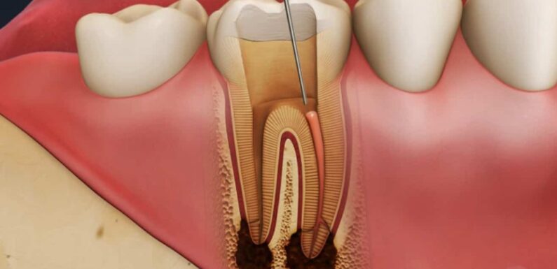
3D imaging provides a three-dimensional view of a patient’s teeth, jaw, and surrounding structures. Unlike standard dental X-rays, which produce flat images, 3D imaging offers a volumetric representation of teeth, allowing endodontists to examine:
- The intricate root canal system
- Bone density and structure
- Areas of infection or inflammation
- Proximity to nerves and sinuses
This detailed view allows an endodontist in Singapore to identify potential complications that may not be visible in traditional 2D X-rays, ensuring a more accurate and safer treatment plan.
Advantages of 3D Imaging for Root Canal Procedures
3D imaging provides several practical benefits during root canal treatments:
1. Precise Diagnosis
Understanding the exact shape, number, and orientation of root canals is critical for successful treatment. Some teeth have multiple or curved canals that may be missed on a standard X-ray. CBCT scans reveal these variations, allowing an endodontist in Singapore to plan the procedure accurately and reduce the risk of incomplete treatment.
2. Enhanced Treatment Planning
With 3D imaging, an endodontist can visualize the full extent of the infection or decay. This information helps determine whether a tooth can be saved through conventional root canal therapy or requires a surgical approach. By mapping out the treatment beforehand, the procedure can be more efficient, reducing discomfort for the patient.
3. Minimizing Radiation Exposure
Although 3D imaging provides detailed information, modern CBCT machines are designed to limit radiation exposure. Compared to multiple conventional X-rays, a single 3D scan delivers precise data with minimal radiation, balancing safety with diagnostic accuracy.
4. Improved Patient Communication
3D images make it easier for patients to understand their dental conditions. By showing a detailed scan of the affected tooth, an endodontist in Singapore can explain the treatment process, potential challenges, and expected outcomes, which helps patients make informed decisions.
How an Endodontist Uses 3D Imaging in Practice
Step 1: Initial Assessment
The process begins with a standard consultation and examination. If the endodontist suspects complex root canal anatomy or hidden infections, a 3D scan is recommended. This scan captures high-resolution images of the teeth and surrounding structures.
Step 2: Planning the Procedure
Using the 3D scan, the endodontist evaluates the tooth’s anatomy in detail. They identify the number of canals, any obstructions, and the location of infection. This step ensures that the treatment plan addresses all factors that could affect the procedure’s success.
Step 3: Performing the Root Canal
During the treatment, the endodontist may refer back to the 3D images to guide precise instrument placement and confirm the removal of infected tissue. The scan also helps prevent accidental damage to nearby nerves or sinuses, which is especially important in teeth located near sensitive structures.
Step 4: Post-Treatment Evaluation
After the procedure, a follow-up scan may be used to verify that the canals are thoroughly cleaned and sealed. This evaluation ensures long-term tooth stability and reduces the likelihood of recurring infection.
Cases Where 3D Imaging is Especially Useful
While not every root canal requires 3D imaging, it is particularly valuable in:
- Teeth with complex or curved canals
- Retreatment cases where previous root canals have failed
- Teeth adjacent to critical nerves or sinuses
- Situations where infection has spread beyond the tooth root
In these cases, the technology allows an endodontist in Singapore to plan and execute treatment with greater precision, reducing procedural risks.
Patient Experience with 3D Imaging
From a patient’s perspective, the process is straightforward and quick. Most modern 3D imaging machines capture the entire scan in less than a minute. Patients remain seated, and the device rotates around their head, producing detailed images almost instantly. The non-invasive nature of the scan and its low radiation dose make it a comfortable and safe option for adults and children alike.
Choosing the Right Endodontist
Selecting an endodontist who uses 3D imaging responsibly ensures that you receive a comprehensive diagnosis and care tailored to your needs. An endodontist in Singapore can interpret the scans effectively and integrate the results into a comprehensive treatment plan, offering precision while minimising risks.
Looking Ahead: The Role of 3D Imaging in Dentistry
3D imaging continues to reshape how endodontists approach complex root canal treatments. By combining imaging technology with specialised skills, it is possible to preserve natural teeth, improve patient outcomes, and reduce complications. Patients benefit from clearer explanations, safer procedures, and more predictable results.
The Clear Picture: Enhancing Dental Care
Incorporating 3D imaging into endodontic treatment represents a shift toward more precise, patient-focused care. For anyone considering a root canal, understanding how an endodontist in Singapore uses this technology can provide reassurance and confidence. With detailed visualisation, careful planning, and attentive care, 3D imaging helps make dental treatments safer and more effective – a clear picture for both patients and practitioners alike.